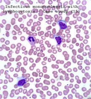
Case study (15)- Infectious mononucleosis with lymphocytosis
History and clinical signs and symptoms
An adolescent girl presented at the outpatient clinic with swollen glands.
She complained about increased pharyngeal pain that intensified when she swallowed.
Other remarkable signs and symptoms included general fatigue, low-grade fever [38.2 °C], swollen glands around the neck, and extensive salivation.
Laboratory Investigation:
1. Hematologic findings:
RBC:4.24 X 10⁶/μL (4-5.5 X 10⁶/μL)
HGB: 133 g/dL (120-174 g/dL)
HCT: 38.3 % (36-52%)
MCV: 90.4 fL (76–96 fL)
MCH: 31.4 pg (27-32 pg)
MCHC: 347 g/L (300-350 g/L)
RDWsd:35.9 fL (20-42 fL)
RDWcv: 14.4 % (0-16 %)
WBC: 11.05 X10³/ μL (5–10 X 103/ μL)
Neutrophils: 4.21 X 10³/μL (2-7.5 X 10³/μL)
Lymphocytes: 4.39 X 10³/μL (1.08–3.17 X 10³/μL)
Monocytes: 1.90 X 10³/μL (0.15-0.7 X 10³/μL)
Eosinophils: 0.06 X 10³/μL (0-0.5 X 10³/μL)
Basophils: 0.50 X 10³/μL (0-0.15 X 10³/μL)
Neutrophils %: 38.1 % (40-75 %)
Lymphocytes %: 39.7 % (14.76-45.4 %)
Monocytes %: 17.2 % (3-7 %)
Eosinophils %: 0.5 % (0-5 %)
Basophils %: 4.5 % (0-1.5 %)
PLT: 124 X 103/μL (150-400 X 103/μL)
Morphological Flags G, Interpretive Flags Basophilia ?, Microcytic PLT?.
Interpretation
Despite the absence of any warning flags, the WBC differential (DIFF) scattergram is not clearly defined. Lymphocyte and monocyte populations are confluent. The WBC count is slightly above the upper limit of the normal range.
A flag „G” point to that immature cells may be present. The basophil (BAS) scattergram indicates a large number of leukocytes.
The lymphocyte, monocyte, and basophil counts and percentages are above the upper limit of the normal range.
The RBC and PLT histograms are well separated and are normal in shape and distribution. The RBC and PLT counts are within the normal range. low MPV value (see interpretive flag “microcytic PLT”) is not clinically relevant here.
Due to the morphologic Flag „G” and the marked increase of basophil cells the assessment of a peripheral blood smear is recommended.
Peripheral blood smear
The percentage of large mononuclear cells presenting the characteristics of monocytes and variant lymphocytes were 6,4% and 29,4% respectively. There is no marked basophilia detected in the peripheral blood smear.
Due to their specific response to lyze reagent immature cells and activated lymphocytes may result in a falsely increased basophil count.
Activated lymphocytes (named as variant forms when evaluated in a microscopic smear) have a similar response to that of monocytes to the effect of lyze reagent. This means they are mistakenly counted as monocytes resulting in a falsely elevated monocyte count with a decreased lymphocyte count.
The analyzer registers a warning flag “G” indicating to the operator that they should conduct a microscopic smear.
2. Other laboratory findings
- Abnormal findings were elevated LDH values (655 U/L [reference range: 230 – 460 U/L); CRP was 61.4 mg/L [<5 mg/L].
Serological tests verified the infection with the Epstein-Barr virus.
Diagnosis
Infectious mononucleosis with lymphocytosis
Disease course
- After 4 weeks on symptomatic therapy, the patient’s general condition improved. A full recovery required several months.


Comments
Post a Comment